Step 1

Pain management

Intranasal

IN fentanyl

2 mcg/kg

Max 100 mcg

Intravenous

IV morphine

0.05 - 0.1 mg/kg

Max 4 mg if <50 kg; max 8 mg if >50 kg

Oral

Ibuprofen

10 mg/kg PO

Max 400 mg

Give within 1 hour of arrival

Step 2

XR: interpreting pediatric elbow injuries in children

Obtain multiple views

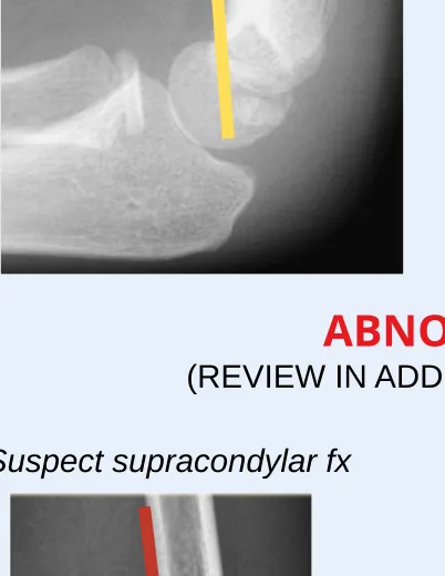
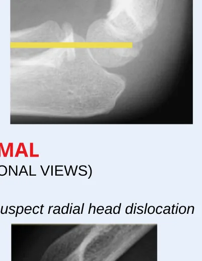
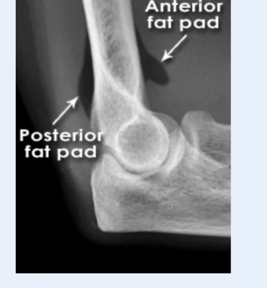
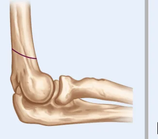
Ensure a true lateral XR before relying on alignment signs.

True lateral elbow radiograph with the figure 8 sign marked.
True lateral Look for the figure 8 sign.
Rotated elbow radiograph labeled not a true lateral.
Not a true lateral Do not anchor on alignment from a rotated lateral.

Alignment

Normal alignment signs only apply when the lateral view is true.

Lateral elbow radiograph with anterior humeral line passing through the middle third of the capitellum.
Anterior humeral line Should pass through the middle third of the capitellum.
Lateral elbow radiograph with radiocapitellar line bisecting the capitellum.
Radiocapitellar line Radial neck should bisect the capitellum.
Abnormal lateral elbow radiograph with anterior humeral line suggesting supracondylar fracture.
Suspect supracondylar fracture Abnormal anterior humeral line warrants review in additional views.
Abnormal lateral elbow radiograph with radiocapitellar line suggesting radial head dislocation.
Suspect radial head dislocation Abnormal radiocapitellar line suggests radial head dislocation.

Fat pads

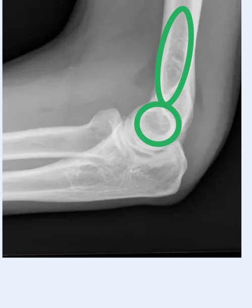
Anterior fat pad can be normal; suspect fracture with anterior sail sign or posterior fat pad.

Elbow radiograph labeled with anterior fat pad and posterior fat pad.
Anterior and posterior fat pads Posterior fat pad or anterior sail sign should raise suspicion for fracture.

Step 3

Gartland classification & local ED management

Type I

Nondisplaced

Illustration of a nondisplaced supracondylar fracture.

Immobilization

  • Posterior long arm splint or cast

Disposition

Discharge for outpatient ortho

Type II

Anterior cortex disrupted

Illustration of a supracondylar fracture with anterior cortex disrupted.

Immobilization

  • Long arm cast
  • Posterior long arm splint

Disposition

Discharge with close ortho follow-up or transfer/admit for OR

Type III

Displaced, anterior and posterior cortex disrupted

Illustration of displaced supracondylar fracture with both cortices disrupted.

Immobilization

  • Posterior long arm splint

Disposition

Transfer/admit for OR

Step 4

Ensure closed and neurovascularly intact

  • Confirm closed injury.
  • Document distal pulses, perfusion, motor function, and sensation.
  • Escalate urgently for neurovascular compromise or open injury.

Source material

Guideline + references

CCMC guideline

Placeholder until Dana/Northwell provides the official guideline URL.

Key references

  1. Teo TL, Schaeffer EK, Habib E, et al. Assessing the reliability of the modified Gartland classification system for extension-type supracondylar humerus fractures. J Child Orthop. 2019;13(6):569-574. doi:10.1302/1863-2548.13.190005
  2. Alton TB, Werner SE, Gee AO. Classifications in Brief: The Gartland Classification of Supracondylar Humerus Fractures. Clin Orthop Relat Res. 2015;473(2):738-741.